
Unilateral Perinephric Pseudocyst in a Young British Shorthair Cat: Diagnostic Imaging and Surgical Outcome
DOI:
https://doi.org/10.71336/ijvar.676Keywords:
Chronic kidney disease, Felis catus, Nephrectomy, Perinephric pseudocyst, UltrasonographyAbstract
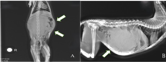
This case report describes the clinical presentation, diagnostic imaging, histopathological findings, and surgical management of a 2-year-old spayed female British Shorthair cat with a unilateral left-sided perinephric pseudocyst. The cat presented with lethargy, anorexia, weight loss, and progressive abdominal distension. Clinical examination revealed a palpable abdominal mass, which was confirmed as a perinephric pseudocyst via ultrasonography and radiography. Surgical intervention involved capsulectomy and nephrectomy, with histopathological confirmation of the diagnosis. The cat initially recovered but developed similar symptoms 54 days postoperatively, with ultrasonographic evidence of cystic structures in the contralateral kidney, necessitating a capsulectomy. Despite initial stabilization, the cat progressed to stage II renal failure 73 days postoperatively and succumbed to the disease. This case highlights that perinephric pseudocyst, although rare, should be considered in the differential diagnosis of young cats presenting with abdominal distension and renomegaly. Notably, biochemical markers of renal pathology may remain within normal limits, underscoring the importance of ultrasonography and histopathology for accurate diagnosis. Although surgical intervention can alleviate symptoms, long-term prognosis remains guarded due to potential recurrence and progressive renal dysfunction. Further studies are needed to explore potential genetic predisposition in British Shorthair cats.
References
Adamama-Moraitou KK, Pardali D, Vafiadis I, Patsikas MN, Prassinos NN. 2018. Perinephric pseudocyst in a cat: management by ultrasound-guided drainage. Journal of the Hellenic Veterinary Medical Society, 68(2): 245. DOI: https://doi.org/10.12681/jhvms.15612
Beck J, Bellenger C, Lamb W, Churcher R, Hunt G, Nicoll R, et al. 2000. Perirenal pseudocysts in 26 cats. Australian Veterinary Journal, 78(3): 166–171. DOI: https://doi.org/10.1111/j.1751-0813.2000.tb10585.x
Debruyn K, Haers H, Combes A, Paepe D, Peremans K, Vanderperren K, et al. 2012. Ultrasonography of the feline kidney. Journal of Feline Medicine and Surgery, 14(11): 794–803. DOI: https://doi.org/10.1177/1098612X12464461
Elliott J, Barber PJ. 1998. Feline chronic renal failure: clinical findings in 80 cases diagnosed between 1992 and 1995. Journal of Small Animal Practice, 39(2): 78–85. DOI: https://doi.org/10.1111/j.1748-5827.1998.tb03598.x
Erdem İ, Zerek A, Cura SE, Yaman M, Deveci MZY, Kırgız Ö. 2022. Exploratory laparotomic diagnosis of renal cystic echinococcosis in a domestic cat from Hatay province of Türkiye and its molecular confirmation. Ankara Üniversitesi Veteriner Fakültesi Dergisi, 69(4): 437-440. DOI: https://doi.org/10.33988/auvfd.1062916
Essman SC, Drost WT, Hoover JP, Lemire TD, Chalman JA. 2000. Imaging of a cat with perirenal pseudocysts. Veterinary Radiology & Ultrasound, 41(4): 329–334. DOI: https://doi.org/10.1111/j.1740-8261.2000.tb02082.x
Griffin S. 2020. Feline abdominal ultrasonography: what’s normal? what’s abnormal? The kidneys and perinephric space. Journal of Feline Medicine and Surgery, 22(5): 409–427. DOI: https://doi.org/10.1177/1098612X20917598
Hickman MA, Cox SR, Mahabir S, Miskell C, Lin J, Bunger A, et al. 2008. Safety, pharmacokinetics and use of the novel NK‐1 receptor antagonist maropitant (Cerenia™) for the prevention of emesis and motion sickness in cats. Journal of Veterinary Pharmacology and Therapeutics, 31(3): 220–229. DOI: https://doi.org/10.1111/j.1365-2885.2008.00952.x
Holloway A, O’Brien R. 2007. Perirenal effusion in dogs and cats with acute renal failure. Veterinary Radiology & Ultrasound, 48(6): 574–579. DOI: https://doi.org/10.1111/j.1740-8261.2007.00300.x
International Renal Interest Society (IRIS). 2023. IRIS Staging of Chronic Kidney Disease. http://www.iris-kidney.com
Kim W, Moon SO, Lee SY, Jang KY, Cho CH, Koh GY, et al. 2006. COMP–Angiopoietin-1 ameliorates renal fibrosis in a unilateral ureteral obstruction model. Journal of the American Society of Nephrology, 17(9): 2474–2483. DOI: https://doi.org/10.1681/ASN.2006020109
Lemire TD, Read WK. 1998. Macroscopic and microscopic characterization of a uriniferous perirenal pseudocyst in a domestic short hair cat. Veterinary Pathology, 35(1): 68–70. DOI: https://doi.org/10.1177/030098589803500107
Mazzanti CM, Castro VSP, Castro JLC, Santalucia S, Raiser AG, Caridade A, et al. 2013. Bilateral perinephric pseudocyst in a cat. Acta Scientiae Veterinariae, 41: 1–5.
McCord K, Steyn PF, Lunn KF. 2008. Unilateral improvement in glomerular filtration rate after permanent drainage of a perinephric pseudocyst in a cat. Journal of Feline Medicine and Surgery, 10(3): 280–283. DOI: https://doi.org/10.1016/j.jfms.2007.11.002
Mouat EE, Mayhew PD, Weh JL, Chapman PS. 2009. Bilateral laparoscopic subtotal perinephric pseudocyst resection in a cat. Journal of Feline Medicine and Surgery, 11(12): 1015–1018. DOI: https://doi.org/10.1016/j.jfms.2009.05.018
Ochoa VB, DiBartola SP, Chew DJ, Westropp J, Carothers M, Biller D. 1999. Perinephric pseudocysts in the cat: a retrospective study and review of the literature. Journal of Veterinary Internal Medicine, 13(1): 47–55. DOI: https://doi.org/10.1111/j.1939-1676.1999.tb02165.x
Placer MA, McManis C. 2019. Laparoscopic resection of bilateral perinephric pseudocyst in a pediatric feline patient. Journal of Feline Medicine and Surgery Open Reports, 5(1): 205511691985064. DOI: https://doi.org/10.1177/2055116919850646
Polzin DJ. 2011. Chronic kidney disease in small animals. Veterinary Clinics of North America: Small Animal Practice, 41(1): 15–30. DOI: https://doi.org/10.1016/j.cvsm.2010.09.004
Salgüero R, Arenas C, Herrtage ME. 2015. Bilateral perinephric pseudocysts in a cat. Veterinary Record Case Reports, 3(1). DOI: https://doi.org/10.1136/vetreccr-2015-000226
Schaefer GDC, Matesco VC, Pereira PR, Panziera W, Driemeier D, Pavarini SP, et al. 2018. Perinephric pseudocyst in a two-month-old female cat. Acta Scientiae Veterinariae, 46(3). DOI: https://doi.org/10.22456/1679-9216.86640
Downloads
Published
How to Cite
Issue
Section
License
Copyright (c) 2026 International Journal of Veterinary and Animal Research (IJVAR)

This work is licensed under a Creative Commons Attribution-NonCommercial-NoDerivatives 4.0 International License.


